"A picture is worth a thousand words."
Dynamic micro-CT
Fast-beating mouse heart
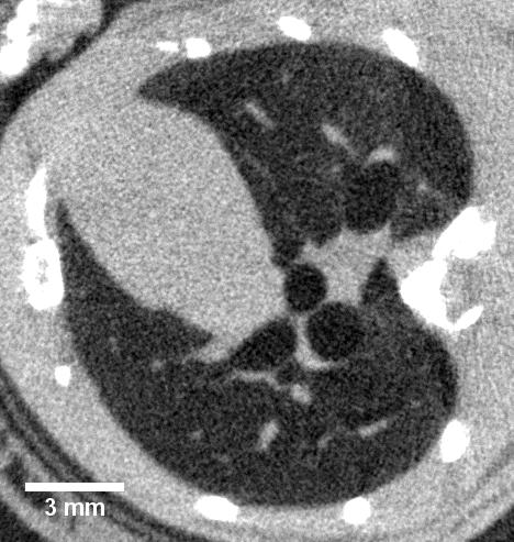
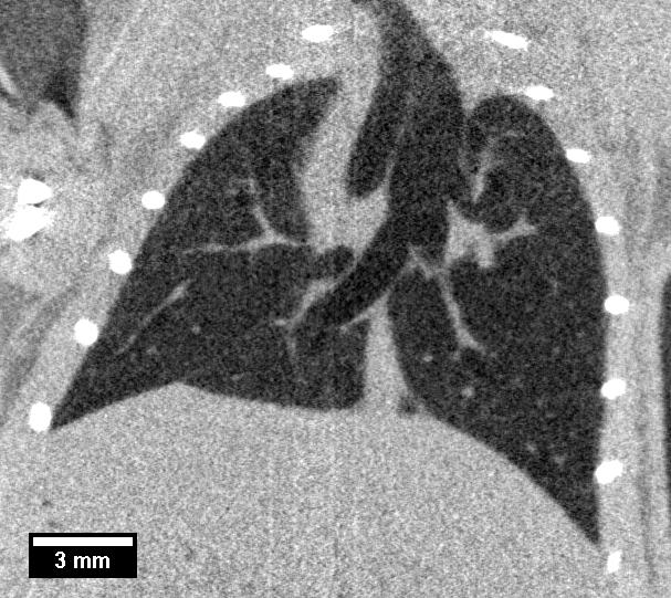
Free-breathing mouse lung
Mouse models of human disease
Heart disease
Lung tumor
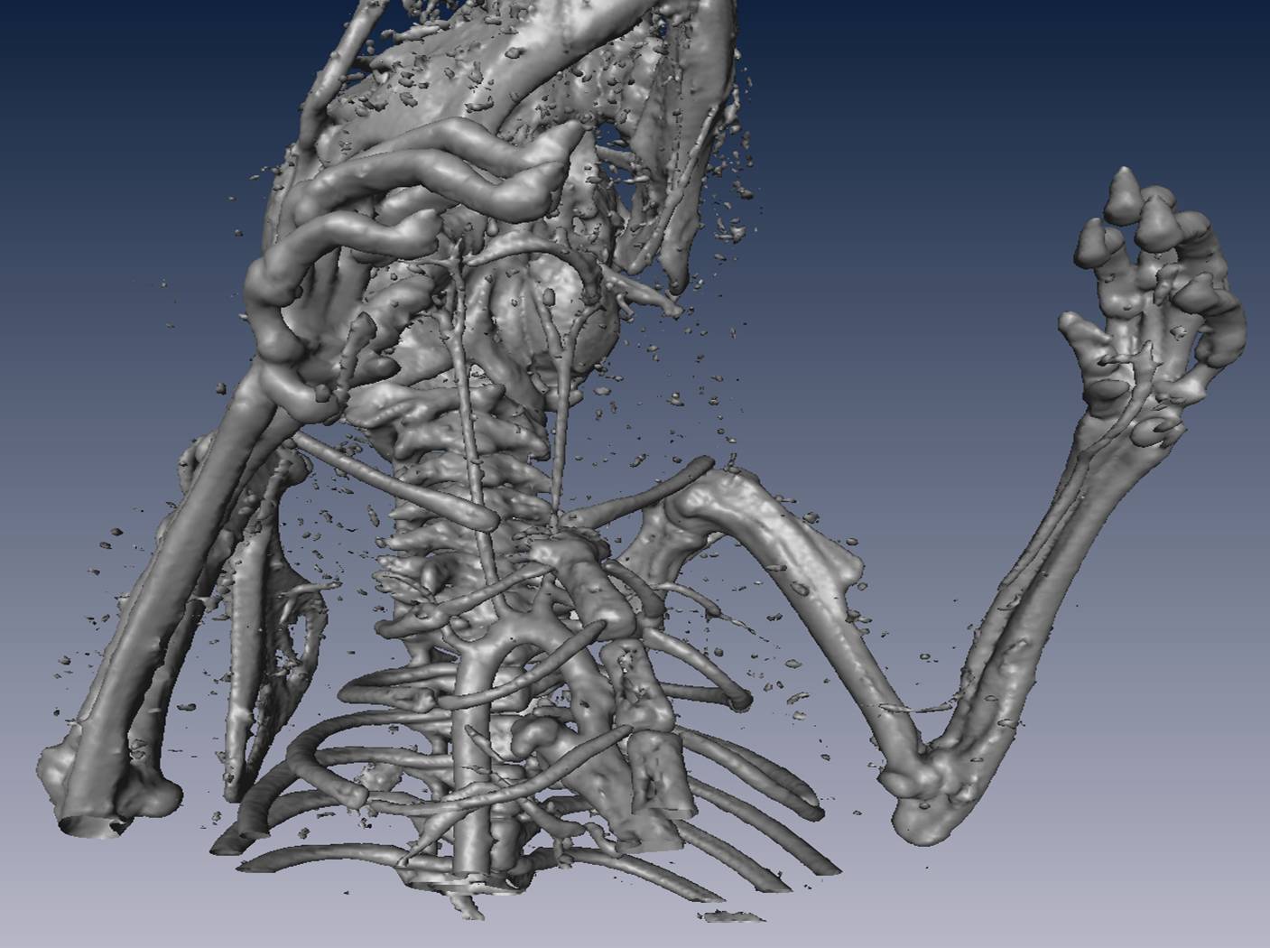
High-resolution micro-CT
Video
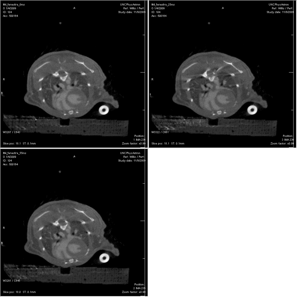
Respiratory and cardiac gated micro-CT imaging of a beating mouse heart under the free-breathing setting. Images were taken at 0ms, 25ms, and 55ms off the R peak from a single C57BL/6 mouse, and data were acquired using a dynamic micro-CT scanner based on a carbon nanotube field emission micro-focus x-ray source.
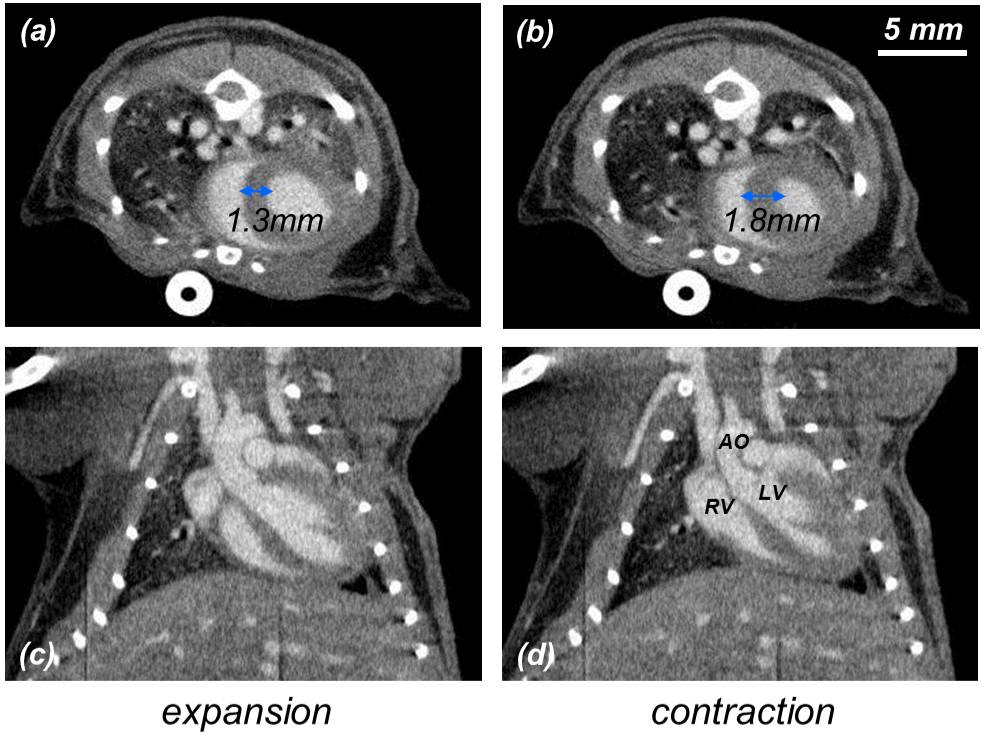
Mouse heart during expansion and contraction. The scanning parameters were 50kVp, 2mA, 15ms pulse width, and 400 views over 200° at a step angle of 0.5°. These images represent 76µm sections taken at the same slice locations from the 3D volumes reconstructed at each time point. The voxel spacing in-plane is also 76µm. The major anatomic structures of the cardiopulmonary vascular system are readily identified in the contrast-enhanced images. All images have the same display window and level. The AO, LV, and RV are labeled for reference.
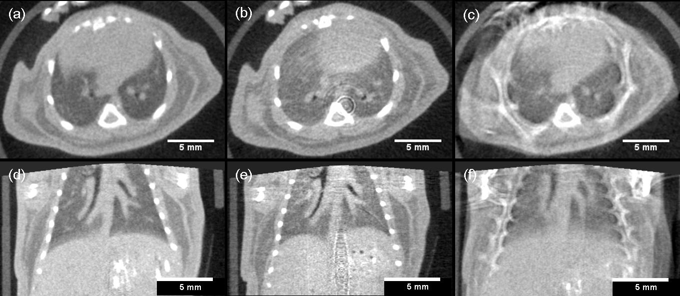
Respiratory-gated micro-CT of free-breathing mice - the very first dynamic CT images from carbon nanotube micro-CT in 2007. The first two columns were gated while the last column was not gated.
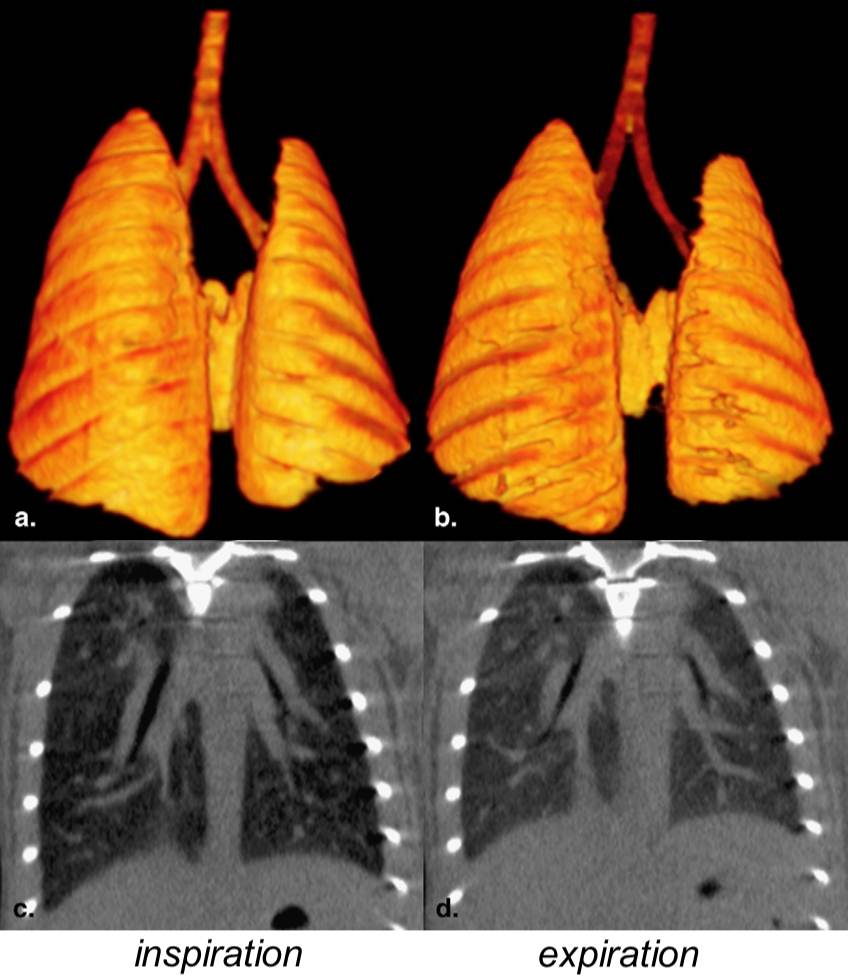
Surface renderings of a mouse lung and trachea in inspiration (a) and expiration (b), and reformatted coronal images of the mouse lung during peak-inspiration (c) and full-expiration (d). Changes in the shape and dimensions of the lungs can be easily appreciated.
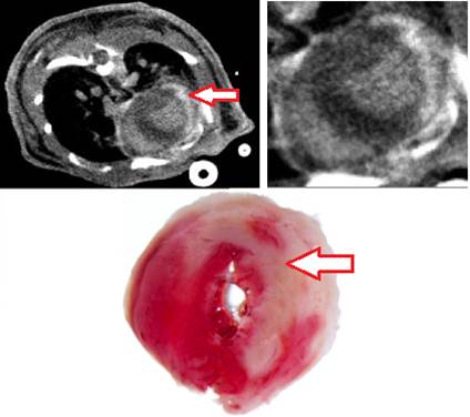
Axial cardiac micro-CT image of the heart of a cardiac ischemia/reperfusion model showing hyper-enhancement (arrow) of the lateral left ventricle wall, representing an area of myocardial infarction. Notice the excellent correlation to histology image at bottom.
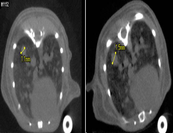
Axial respiratory gated micro-CT image of the lower thorax of mouse model of multifocual lung tumors. The left image was taken at the 1st week showing a largest lesion of 1.1mm at the right lung base (left of the image). The right image was taken at the 3rd week from the same mouse showing the lesion has grown to 1.5mm.


